Why Renal Ultrasound Is the First Step When Kidneys Don’t Look Right
If your doctor suspects a blockage in your urinary tract or thinks your kidneys might be swollen, shrinking, or damaged, they’ll almost always start with a renal ultrasound. It’s quick, safe, and doesn’t use radiation. Unlike CT scans or MRIs, you don’t need to fast, drink contrast dye, or lie in a tight tube. You just lie on your back, and a tech moves a small device over your sides and belly. In under 30 minutes, they can tell if your kidneys are the right size, if urine is backing up, or if there’s something blocking the flow.
What Exactly Does a Renal Ultrasound Show?
A renal ultrasound doesn’t just take a picture-it measures. The tech checks several key numbers:
- Kidney length: Normal adult kidneys are 9 to 13 centimeters long. If one is significantly shorter, it could mean long-term damage or scarring.
- Cortical thickness: The outer layer of the kidney should be at least 1 cm thick. Thinning here often signals chronic kidney disease.
- Renal pelvis diameter: The space where urine collects before flowing to the bladder. If it’s wider than 7 mm, it’s called hydronephrosis-meaning urine is backing up.
- Resistive index (RI): This is a Doppler measurement that shows how easily blood flows through the kidney’s arteries. An RI of 0.70 or higher strongly suggests obstruction. Studies show this number is 87% accurate at catching blockages.
These aren’t guesses. They’re based on decades of clinical data and standardized protocols. When all these numbers line up-like a kidney that’s enlarged with a dilated pelvis and an RI above 0.70-it’s a clear sign something’s blocking urine flow.
How Ultrasound Finds Obstruction-Even When You Can’t See It
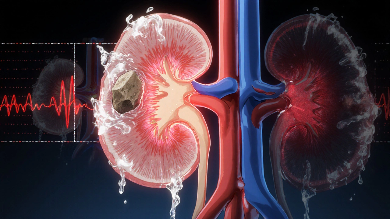
Obstruction doesn’t always mean a big stone. It could be a narrow ureter, a twisted ureteropelvic junction (UPJ), or even a tumor pressing from outside. Ultrasound catches these by looking at the plumbing system inside the kidney. When urine can’t drain, pressure builds. That pressure stretches the renal pelvis and calyces-the tiny chambers where urine collects. This swelling is called hydronephrosis.
Doctors grade hydronephrosis using the Society for Fetal Urology system: mild, moderate, or severe. Mild might just be a slightly widened pelvis. Severe means the whole inner structure is ballooned, and the kidney tissue is starting to thin out. That’s when things get serious-because prolonged pressure can permanently damage kidney function.
Advanced Doppler techniques now help spot hidden causes. For example, a blood vessel crossing over the ureter (called a crossing vessel) can compress it and cause UPJ obstruction. Doppler ultrasound can detect the altered blood flow patterns around that spot. Newer tools like shear-wave elastography even measure how stiff the kidney tissue has become. The more pressure from backup urine, the stiffer the kidney gets. This isn’t standard everywhere yet, but it’s showing up in research hospitals and could become routine soon.
Ultrasound vs. CT vs. MRI: Why Ultrasound Still Wins for Initial Checks
People often think CT scans are the gold standard-and they are, for finding tiny stones. But here’s the catch: a single CT scan gives you about 10 millisieverts of radiation. That’s like 500 chest X-rays. For someone with recurrent kidney stones, that adds up fast.
Ultrasound finds about 80% of stones larger than 3 mm. It won’t catch a 1-mm stone like a CT can, but for most cases, that’s enough. More importantly, it tells you if the stone is causing a backup. A CT might show a stone and nothing else. Ultrasound shows the stone and the swollen kidney behind it.
MRIs give better soft tissue detail but cost 3 to 5 times more-up to $2,500-and still can’t reliably see stones. Nuclear scans show how well the kidney is working but use radiation and give fuzzy images. Ultrasound? Around $300. No radiation. No contrast. No waiting. It’s the obvious first step.
Emergency departments use point-of-care ultrasound to cut diagnosis time by nearly an hour. Instead of waiting for a radiologist to schedule a formal scan, the ER doctor can do it right at the bedside. If the kidney looks swollen and the RI is high, they know it’s obstruction and can act fast.
When Ultrasound Falls Short-and What Comes Next
Ultrasound isn’t perfect. It struggles with very large patients. If your BMI is over 35, sound waves can’t penetrate deep enough. The images get blurry, and measurements become unreliable. In those cases, doctors move to CT or MRI.
Another limitation? Operator skill. A 2018 study found up to 20% variation in kidney size measurements between inexperienced and expert sonographers. That’s why training matters. The American Institute of Ultrasound in Medicine recommends at least 40 supervised exams before someone is considered competent. Many radiology residents say it takes about 50 cases to feel confident measuring resistive index accurately.
Also, ultrasound can’t tell you how fast urine is draining. That’s something CT with special 3D algorithms can do-but it’s not widely available. If your doctor suspects a slow-draining kidney, they might still order a nuclear scan (called a MAG3 renogram) to measure drainage rate, even after a normal ultrasound.
What Happens After the Scan? How Doctors Use the Results
Results aren’t just numbers. They’re decisions.
- If your kidney is normal size, cortex is thick, pelvis is under 7 mm, and RI is below 0.70-you’re likely fine. No further imaging needed.
- If you have mild hydronephrosis and no pain? Your doctor might watch and repeat the ultrasound in 6 to 12 weeks.
- If you have severe hydronephrosis, high RI, and a stone? You’re likely headed for a urologist. You may need a stent placed to bypass the blockage or surgery to fix a narrowed UPJ.
- If you’re pregnant? Ultrasound is the only safe option. It’s the go-to tool for checking kidney swelling in pregnancy-related hydronephrosis.
- If you’re a child with a history of urinary tract infections? Ultrasound is the standard to check for congenital obstructions like UPJ stenosis.
For patients with known blockages-like after UPJ repair surgery-doctors often use weekly or monthly ultrasounds to track progress. No radiation. No cost. No hassle. That’s why one urologist told a medical forum: “I track hydronephrosis weekly in post-op UPJ patients with bedside ultrasound instead of exposing them to repeated radiation.”

The Future: AI, Super-Resolution, and Quantitative Ultrasound
Ultrasound isn’t standing still. Researchers are now using artificial intelligence to automatically grade hydronephrosis. Mayo Clinic is testing AI tools that can measure kidney swelling with the same accuracy as a radiologist-faster and without human error.
Super-resolution ultrasound is another breakthrough. It can now detect tiny blood vessels in the kidney cortex. In the future, this might let doctors see early signs of scarring or reduced blood flow before the kidney even starts to shrink.
And then there’s ultrasound localization microscopy (ULM). It’s still experimental, but it could one day count individual nephrons-the filtering units of the kidney. Imagine being able to track how many nephrons you’ve lost over time, without a biopsy. That’s the direction we’re heading.
What You Need to Know Before Your Scan
You don’t need to fast. You don’t need to stop your meds. Just drink a glass of water an hour before. A full bladder helps push bowel gas out of the way and gives a clearer view of the lower ureters and bladder.
The scan itself is painless. You’ll feel a little cold gel and pressure from the probe. No needles. No shocks. No noise beyond a soft beep from the machine.
Results usually come back within 24 to 48 hours. If your doctor says “your kidneys look good,” take it seriously. If they say “there’s some swelling,” don’t panic. Ask: “What’s the grade? What’s the resistive index? Is it on one side or both?” Those numbers tell the real story.
Can renal ultrasound detect kidney stones?
Yes, but not all of them. Renal ultrasound detects about 80% of stones larger than 3 mm. Smaller stones (under 2 mm) are often missed. CT scans are better for finding tiny stones, but ultrasound is preferred for initial checks because it shows if the stone is causing a backup-something CT alone can’t tell you.
Is renal ultrasound safe during pregnancy?
Yes, it’s the safest and most commonly used imaging test during pregnancy. It uses no radiation or contrast agents, making it ideal for monitoring kidney swelling, which is common in pregnant women due to hormonal changes and pressure from the growing uterus.
What does a high resistive index mean?
A resistive index (RI) of 0.70 or higher suggests increased resistance to blood flow in the kidney’s arteries, often caused by urine backup from obstruction. Studies show this measurement has 87% sensitivity and 90% specificity for detecting obstructive uropathy. It’s one of the most reliable ultrasound signs of blockage.
Can obesity affect the accuracy of a renal ultrasound?
Yes. When a patient’s BMI exceeds 35, sound waves have trouble penetrating deep enough to get clear images of the kidneys. In these cases, the ultrasound may be inconclusive, and doctors often turn to CT or MRI for better visualization.
How long does a renal ultrasound take?
Typically 15 to 30 minutes. No special preparation is needed beyond drinking water to fill your bladder. The procedure is painless, and results are usually available within 1 to 2 days.
Do I need a referral for a renal ultrasound?
Yes. In most healthcare systems, you need a referral from a doctor or specialist. It’s not a screening test for healthy people-it’s used when there’s a clinical suspicion of kidney obstruction, size changes, or other kidney disease.
Can renal ultrasound detect kidney cancer?
It can detect masses or abnormal growths in the kidney, but it can’t always tell if they’re cancerous. A suspicious mass seen on ultrasound usually requires follow-up with CT or MRI for better characterization. Ultrasound is a good first step, but not definitive for cancer diagnosis.
What to Do After Your Ultrasound Results
If your scan is normal, no action is needed unless symptoms return. Keep drinking water, watch for pain or fever, and follow up if you notice changes in urine color or output.
If you have hydronephrosis, ask your doctor: “Is it mild, moderate, or severe? Is it getting worse? What’s the next step?” Don’t assume it’s just a stone. It could be a structural issue needing surgery, or a sign of something more serious like a tumor.
For people with chronic kidney disease or recurrent blockages, regular ultrasounds are a lifeline. They’re the only imaging tool you can safely use every few months without risking radiation damage. That’s why they’re used in dialysis centers, transplant clinics, and pediatric nephrology units around the world.
Renal ultrasound isn’t flashy. But in the real world of patient care, it’s the quiet hero that catches problems early, avoids unnecessary radiation, and saves lives one scan at a time.
Comments
Erika Sta. Maria
okay but like… if ultrasound is so great why do we still have CTs? it’s 2024 and we’re still using a 1950s tech because it’s ‘cheap’? 🤡 i’ve had 3 kidney scans and 2 of them were inconclusive because my BMI is 32 and the tech said ‘we can’t see past the fat’ - so now i’m stuck with radiation just to get a straight answer. also, resistive index? that’s not even a real number, it’s a guess wrapped in jargon. my nephrologist admitted he ‘eyeballs’ it half the time. stop pretending this is science.
Nikhil Purohit
bro this is actually one of the clearest breakdowns of renal ultrasound i’ve ever read. i’m a med student in delhi and we barely get taught this in depth - we just memorize ‘hydronephrosis = bad’ and move on. the part about cortical thickness and RI being 87% accurate? that’s gold. i’m printing this out and taping it to my desk. also, the point about pregnant patients? huge. my aunt had hydronephrosis during her third trimester and they did 4 ultrasounds - zero radiation, zero drama. this is how medicine should work.
Debanjan Banerjee
Let me address the misinformation in the previous comment. The resistive index is not an ‘eyeballed’ metric - it is a validated Doppler-derived parameter with interobserver reliability coefficients exceeding 0.85 in peer-reviewed studies (J Ultrasound Med, 2020). The claim that ultrasound fails in BMI >32 is misleading: while image quality degrades, experienced sonographers still achieve diagnostic accuracy in 70–80% of cases using harmonic imaging and lower-frequency transducers. Furthermore, the 80% stone detection rate is not a flaw - it’s a feature. The goal is not to find every stone, but to identify obstruction, which ultrasound does with greater clinical context than CT. CT finds stones; ultrasound finds dysfunction. That’s why it’s first-line. And yes, it’s cheaper - but more importantly, it’s safer, faster, and repeatable. This isn’t 1950s tech - it’s precision medicine with a 40-year evidence base. Stop romanticizing radiation.
Shawn Sakura
just wanted to say THANK YOU for writing this. as a dad whose kid had a UPJ obstruction at 3 months old, this is the exact info i wish i’d had when we were freaking out in the hospital. we did 7 ultrasounds in 6 months - no needles, no scary machines, just quiet beeps and a tech saying ‘looks better today.’ i cried the first time the pelvis dropped below 7mm. this isn’t just tech - it’s hope. keep sharing this stuff. 🙏
Paula Jane Butterfield
One sentence: ultrasound is the quiet superhero of nephrology - no cape, no fanfare, just reliable, safe, and everywhere it’s needed.